pEYFP-Golgi
- 产品货号GS-1123
- 销售价格¥ 680
- 包装规格2μg质粒或0.5ml甘油菌
- 载体抗性Kanamycin (卡那霉素)
- 筛选标记新霉素(Neomycin)
- 载体大小4900 bp
- 启动子CMV
载体说明
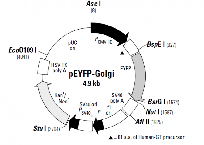
pEYFP-Golgi encodes a fusion protein consisting of enhanced yellow fluorescent protein (EYFP) and a sequence encoding the N-terminal 81 amino acids of human beta 1,4-galactosyltransferase (GT; 1). This region of human beta 1,4-GT contains the membrane-anchoring signal peptide that targets the fusion protein to the trans-medial region of the Golgi apparatus (2–4). The EYFP gene contains the four amino acid substitutions previously published as GFP-10C (5). The fluorescence excitation maximum of EYFP is 513 nm; the emission spectrum has a peak at 527 nm (in the yellow-green region). When excited at 514 nm, the Emof EYFP is 84,500 cm–1M–1 and the fluorescence quantum yield is 0.61 (6), resulting in a bright fluorescent signal. The fluorescence observed is roughly equivalent to that of EGFP.
In addition to the chromophore mutations, EYFP contains more than 190 silent base changes that correspond to human codon-usage preferences (6). SV40 polyadenylation signals downstream of the EYFP-Golgi fusion direct proper processing of the 3' end of the mRNA. The vector backbone also contains an SV40 origin for replication in mammalian cells expressing the SV40 T-antigen. A neomycin resistance cassette (Neor) consisting of the SV40 early promoter, the neomycin/ kanamycin resistance gene of Tn5, and polyadenylation signals from the herpes simplex virus thymidine kinase (HSV-TK) gene allow stably transfected eukaryotic cells to be selected using G418 (7). A bacterial promoter upstream of this cassette drives expression of the gene encoding kanamycin resistance in E. coli. The pEYFP-Golgi backbone also provides a pUC origin of replication for propagation in E. coli and an f1 origin for single-stranded DNA production.
载体应用
The pEYFP-Golgi Vector is designed for fluorescent labeling of the trans-medial region of the Golgi apparatus in mammalian cells. Fluorescence can be observed in living cells by microscopy or flow cytometry. pEYFP-ER can be introduced into mammalian cells using any standard transfection method. If required, stable transformants can be selected using G418 (7). Filter sets are available for dual-color detection of EYFP and ECFP using conventional epifluoresence microscopy (8). Please refer to the Living Colors? User Manual, provided with this vector, for additional information.